A chondroma is a benign tumor that develops in the cartilage tissue. It can occur anywhere in the body where cartilage is present. However, it is most commonly found in the bones of the hands and feet.
Chondroma Symptoms
Pain
Pain is the most common symptom of chondroma. It may be a relatively dull ache or a sharp, shooting pain that increases with activity.
Swelling
Chondromas can cause swelling in the affected area.
Weakness
Chondromas that develop in the bones of the hands or feet can cause weakness in the affected area.
Numbness or tingling
Chondromas that develop near nerves can cause numbness or tingling in the affected area.
Also From Science Reflex:
- What are Bedoyecta Injections?
- What Are Critical Health Supplements?
- What Is Groin Massage? How Can It Benefit You?
Chondroma Treatment
While they are generally not cancerous, they can cause discomfort and damage to the surrounding tissues.
Here are some standard treatment options for chondroma.
Observation
In some cases, small and asymptomatic chondromas can be observed without any treatment. Your doctor may monitor the tumor through regular imaging tests to make sure it is not growing or causing any issues.
Medications
Pain relievers may be prescribed to manage chondroma.
Physical therapy
Physical therapy may be recommended to help improve the range of motion and strengthen muscles around the affected area.
Surgery
If the chondroma is causing significant pain, swelling, or functional impairment, surgery may be recommended to remove the tumor.
Juxtacortical Chondroma
Juxtacortical chondroma is a rare type of benign bone tumor that develops close to the cortex of a bone.
It is typically slow-growing and does not spread to other parts of the body.
Causes of juxtacortical chondroma
The exact cause of the juxtacortical chondroma is unknown. It may be caused by trauma or injury to the bone, or it may be related to genetics.
Symptoms of juxtacortical chondroma
In many cases, juxtacortical chondromas do not show any symptoms and are discovered incidentally during imaging tests for other conditions.
Diagnosis
Juxtacortical chondromas can be diagnosed through imaging tests such as X-rays, MRI, or CT scans.
Treatment
The treatment of juxtacortical chondroma depends on the size and location of the tumor. The patient’s overall health is also a factor.
Small tumors that are not causing any symptoms may be observed without any treatment.
Benign Chondroma
Benign chondroma is a non-cancerous tumor that develops in the cartilage tissue.
They can appear in any part of the body where cartilage is present, including the bones, joints, and soft tissues.
Causes of benign chondroma
The exact cause of benign chondroma is unknown, but it may be related to genetic mutations or abnormalities in the cartilage cells.
Symptoms of benign chondroma
In many cases, benign chondromas do not cause symptoms and are discovered incidentally during imaging tests for other conditions.
However, if the tumor grows bigger, it may cause pain, swelling, or discomfort in the affected area.
Diagnosis of benign chondroma
Benign chondromas can be diagnosed through imaging tests such as X-rays, MRI, or CT scans.
Treatment
The treatment of benign chondroma depends on the size, location, and severity of the tumor, as well as the patient’s overall health.
Cortical Chondroma
Cortical chondroma is a rare type of benign bone tumor that arises from the cartilage cells within the cortex, or outer layer, of the bone.
This tumor is most commonly found in long bones – femur, tibia, and humerus.
Symptoms of cortical chondroma
The most common symptom of cortical chondroma is pain, which can be localized to the affected bone or may radiate to other areas.
Diagnosis
Cortical chondromas are typically diagnosed using imaging tests, including X-rays, CT scans, and MRI scans.
Treatment
Although this tumor can cause pain and other symptoms, it is generally slow-growing and has a good success rate with surgical removal.
Regular follow-up visits with your healthcare provider are necessary to monitor for any signs of recurrence or complications.
Parosteal Chondroma
Parosteal chondroma is a benign bone tumor. It grows from the cartilage cells and usually occurs in the surface layer of the bone.
Symptoms of parosteal chondroma
Parosteal chondromas are often asymptomatic and are discovered incidentally on X-rays.
However, some patients may experience pain, swelling, or a visible lump in the affected area.
Diagnosis
The diagnosis of parosteal chondroma is based on the patient’s history, physical examination, and radiographic imaging.
Treatment
Surgical removal is the standard treatment for parosteal chondroma.
Pulmonary Chondroma
Pulmonary chondroma is a rare benign tumor. It arises from the cartilage cells within the lung.
Symptoms
In rare cases, patients may experience cough, chest pain, shortness of breath, or coughing up blood.
Diagnosis
The diagnosis of pulmonary chondroma is usually based on radiographic imaging studies such as chest X-rays, CT scans, or MRI scans.
Treatment
Surgical resection is the most popular treatment for pulmonary chondroma.
Condylar Chondroma
Condylar chondroma arises from the cartilage cells of the temporomandibular joint (TMJ). Its most common symptoms include pain, swelling, and limited jaw movement.
The diagnosis of condylar chondroma is usually based on clinical symptoms, radiographic imaging studies, and histopathological examination.
Surgical resection is done for phytologic chondroma.
Phytologic Chondroma
Phytochondroma, also known as physeal chondroma, is a rare benign tumor that arises from the cartilage cells within the growth plate of long bones.
The most common symptoms of phytologic chondroma include the following:
- Pain
- Swelling
- Limited range of motion.
The diagnosis of phytologic chondroma is usually based on clinical symptoms, radiographic imaging studies, and histopathological examination.
These chondromas are removed surgically.
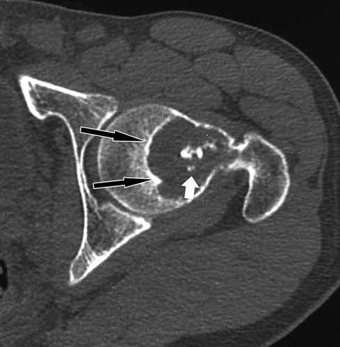
Pelvic Chondroma
Pelvic chondroma is a rare benign tumor that arises from the cartilage cells within the pelvis. Its most common symptoms of pelvic chondroma include:
- Pain
- Swelling
- Limited range of motion in the affected area.
Pelvic chondroma is usually diagnosed based on clinical symptoms, radiographic imaging studies, and histopathological examination.
It can also be removed surgically.